
Tracking How T Cells Evolve in Graft-Versus-Host Disease
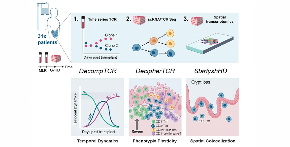
A new study from the Azizi and Reshef labs provides a detailed look at how immune cells evolve and contribute to tissue damage in graft-versus-host disease (GVHD), a serious complication that can occur following bone marrow and blood stem-cell transplantation. The study provides a new way to connect what immune cells are doing in the blood with where they ultimately cause tissue damage. Eventually, this insight can help researchers develop approaches for earlier prediction and more targeted treatment of GVHD.
Published in Nature Immunology, “Spatiotemporal single-cell profiling reveals T cell clonal dynamics and phenotypic plasticity in human graft-versus-host disease” includes several IICD researchers from the Azizi and McFaline-Figueroa groups among its authors.
Understanding Graft-Versus-Host Disease
In GVHD, immune cells from the donor, particularly T cells, recognize the patient’s tissues as foreign and attack them. While researchers understand that T cells drive damage, they have had difficulty determining which individual clones are involved, how they change over time, and what happens after they enter damaged tissue.
To learn more about the process, the team generated a single-cell atlas of human GVHD directly from patient intestinal tissue samples, providing a detailed view of immune-cell states in affected tissue.
“Each T cell carries a unique T-cell receptor sequence that acts like a molecular barcode, allowing us to identify donor-reactive T-cell clones before transplantation and then track those same clones in patients over time,” Dr. Lingting Shi, one of the study’s leading authors, explains. “A combination of methods allowed us to determine what states these cells adopt in the gut, where they are located in intestinal tissue, and whether T-cell populations originated from the donor or the recipient.”
Tracking T Cells Across Time and Space
To connect different dimensions of the immune response, the researchers developed and adapted computational tools that showed how T-cells clones expand, change, and localize in damaged tissues.
This study integrated clinical transplantation, immunology, single-cell genomics, spatial transcriptomics, and computational modeling. Beyond its biological findings, the study also provides a computational resource: researchers developed DecompTCR for longitudinal clonal dynamics and adapted DecipherTCR and StarfyshHD to connect clonal phenotype with tissue localization. The framework provides a way to study immune-cell behavior across multiple scales—from an individual T-cell clone to an entire tissue microenvironment.
“The broader significance is that we move from a static picture of GVHD toward a spatiotemporal and lineage-resolved view of disease,” Ajna Uzuni, one of the study’s leading authors, reflects. “We can ask not only which immune cells are present, but where they came from, which clones they belong to, how they evolved, where they moved, and where they ultimately caused damage.”
Connecting T Cells to Intestinal Damage
From the study, researchers found that in severe GVHD, donor-reactive T-cell clones were more likely to undergo persistent expansion, particularly among CD8 T cells.
They also examined the effects of post-transplant cyclophosphamide, a treatment used to help prevent GVHD. The researchers found that the treatment preferentially removes rapidly expanding donor-reactive T-cell clones, suggesting that clones that expand less before treatment may be more likely to remain after treatment and later contribute to severe disease.
“Once donor-derived CD8 T-cell clones entered the intestine, they were not static,” shares Dr. Ximi K. Wang, one of the study’s leading authors. “The same clones could acquire a tissue-resident memory program as they moved toward the intestinal lining, while retaining their ability to kill cells.”
Spatial analysis showed that these cells accumulated near the bases of intestinal crypts, which contain the stem cells responsible for continually regenerating the intestinal lining. They were particularly enriched in regions where crypts had been damaged or lost.
Toward Earlier Detection and New Therapeutic Strategies
The study could inform future approaches to identifying and treating GVHD. Tracking the behavior of alloreactive T-cell clones in the blood may eventually provide an early indicator of a patient’s risk of developing severe disease.
Similarly, examining how immune cells are organized within intestinal tissue could offer a tissue-level biomarker, showing the extent of tissue damage or a patient’s response to treatment. However, these applications will require validation in larger patient cohorts before they can be applied clinically.
The researchers also identified immune-cell populations and cellular programs that could provide directions for future therapeutic research. More broadly, the computational framework developed through the study could be applied beyond GVHD to other conditions in which persistent T-cell clones play an important role, including transplantation, autoimmune disease, infection, and cancer.
Next Steps
Researchers identified three potential directions for future research: validating the findings in larger and more diverse transplant cohorts; understanding what causes CD8 T cells to acquire the tissue-resident program and whether that transition can be therapeutically interrupted; and investigating how immune cells converge around intestinal stem-cell niches.
“The spatial findings motivate us to understand the multicellular interactions surrounding intestinal stem-cell niches: why certain immune cells converge there, how those interactions contribute to crypt destruction and failed tissue repair, and whether disrupting those pathological cellular neighborhoods could protect the intestine,” concludes Dr. Shi.
Publication Details
The paper was published in Nature Immunology in September 2026 (DOI: 10.1038/s41590-026-02631-2). The work was a collaboration between the groups of Elham Azizi (Irving Institute for Cancer Dynamics and Department of Biomedical Engineering) and Ran Reshef (Professor of Medicine). The study was led by Lingting Shi (IICD), Ajna Uzuni, and Ximi K. Wang.